Human Cerebellar Organoid Platform
Engineered 3D human cerebellar organoids recapitulating cerebellar cytoarchitecture, including Purkinje cells, granule cells, and functional GABAergic microcircuits. Optimized for modeling spinocerebellar ataxias, neurodevelopmental disorders, and targeted cerebellar drug screening.
Cerebellar Cytoarchitecture & Functional Circuitry
Our human cerebellar organoids accurately model embryonic hindbrain patterning, delivering polarized cell layers and synaptic networks crucial for studying motor coordination and cerebellar pathologies.
Purkinje Cell Differentiation
Generates mature PCP2+ and Calbindin+ Purkinje neurons with extensive dendritic arborization and characteristic spontaneous action potential firing patterns.
Granule & Interneuron Networks
Incorporates ATH1+ cerebellar granule cell progenitor populations and VGAT+ GABAergic interneurons, enabling physiologically relevant synaptic microcircuits.
Midbrain-Hindbrain Boundary
Patterned using precise FGF8 and WNT signaling gradients to recreate the isthmic organizer profile essential for authentic cerebellar regional identity.
Standardized Differentiation Protocol
A stringently validated 4-stage differentiation pathway engineered to yield uniform cerebellar organoids with consistent neuronal lineage proportions.
Spheroid Induction
hiPSCs are aggregated into uniform neurospheroids using ultra-low attachment microplates under defined conditions.
Isthmic Patterning
Application of FGF8b and GDF7 morphogens directs neural progenitors toward cerebellar and hindbrain lineages.
Layer Stratification
Bioreactor cultivation fosters Purkinje cell maturation, dendritic arbor development, and granule cell migration.
Quality Control
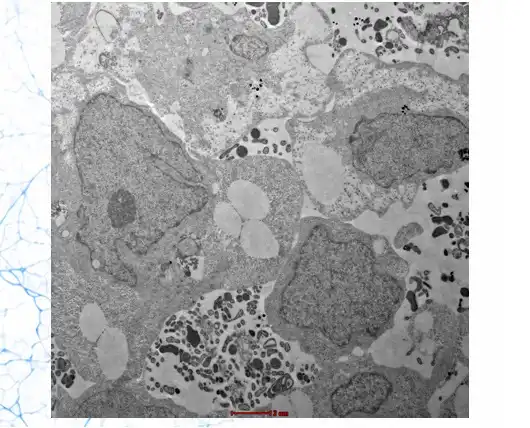
Validation via Calbindin/PCP2 immunostaining, MEA electrophysiology, structural uniformity, and sterility verification.
Technical Profile & QC Standards
| Cell Source | Control (Male/Female) or Disease-Specific hiPSC Lines |
| Organoid Diameter | 1.8 mm ± 0.2 mm (Standardized at Day 60 maturation) |
| Plate Formats | 96-well / 384-well ULA Plates or Custom Assay Assortments |
| Key Markers | Calbindin, PCP2, EN2, BARHL1, PAX6, VGAT, GFAP |
| Functional Validation | MEA verified Purkinje action potential firing & GABAergic tone |
| Quality Assurance | Mycoplasma Negative, Sterility Verified, >90% Viability |
| Shipment Options | Active temperature-controlled media or Cryopreserved vials |
Translational Research Applications
Bridging the gap in cerebellar drug discovery, toxicity screening, and genetic disease therapeutic testing.
Spinocerebellar Ataxia (SCA) Modeling
Utilize patient-derived or CRISPR-edited iPSC variants to model polyglutamine expansion diseases (SCA1, SCA2, SCA3) and test antisense oligonucleotide (ASO) or small molecule therapies.
Autism Spectrum & Neurodevelopmental Research
Investigate cerebellar circuit dysregulation and granule cell migration defects linked to neurodevelopmental conditions such as ASD and tuberous sclerosis.
Cerebellar Toxicity & Drug Safety Screening
Evaluate drug candidate neurotoxicity specifically targeting Purkinje cells or cerebellar network electrophysiology in high-throughput microplate formats.
Medulloblastoma & Tumor Microenvironment Studies
Co-culture primary medulloblastoma cells with human cerebellar organoids to study tumor initiation, invasion kinetics, and targeted oncological treatments.
Frequently Asked Questions
Common queries regarding cerebellar organoid culture, shipment, and experimental setup.