Neural Progenitor Biology
Investigate neural progenitor populations, differentiation processes, and developmental changes within a three-dimensional neural environment.
Human stem-cell-derived three-dimensional neural models designed to support research into early brain development, developmental neurobiology, disease mechanisms, and translational neuroscience.
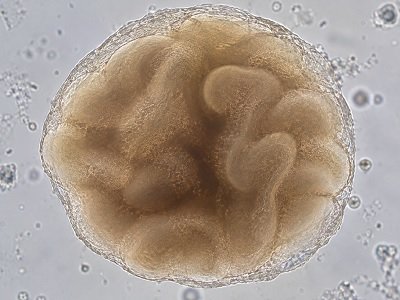
Fetal brain organoid models provide a three-dimensional research environment for investigating selected aspects of early human neural development.
Human brain development involves dynamic interactions between neural progenitors, differentiating cells, extracellular environments, and emerging tissue architecture.
Fetal brain organoids provide a complementary model system in which researchers can investigate developmental processes within a three-dimensional cellular environment.
These models can support research across developmental neuroscience, neurobiology, disease modelling, drug research, and advanced cellular imaging.
Fetal brain organoids can provide a useful experimental context for studying cellular and structural events associated with early human brain development.
Investigate neural progenitor populations, differentiation processes, and developmental changes within a three-dimensional neural environment.
Examine spatial cellular organization and tissue-level features that are difficult to capture in conventional two-dimensional culture systems.
Support research into differentiation trajectories and changes associated with neural maturation in organoid culture.
Explore different neural and supporting cell populations within a complex three-dimensional model.
Investigate experimental perturbations affecting developmental signalling, differentiation, and cellular organization.
Combine microscopy and cellular assays to characterize morphology, organization, and experimental phenotypes.
Organoid models can be integrated with complementary analytical approaches to investigate changes across developmental states.
Study processes associated with neural progenitor activity, differentiation, cellular organization, and early tissue patterning in a controlled experimental model.
Compare control and experimental conditions to investigate how genetic, chemical, environmental, or pharmacological changes influence developmental phenotypes.
A structured workflow can help researchers connect organoid culture with imaging, molecular characterization, and downstream analysis.
Establish and maintain the selected human neural organoid model under the defined research protocol.
Monitor morphology, culture condition, developmental progression, and experimental consistency.
Apply appropriate imaging, cellular, molecular, or functional assays according to the research objective.
Compare experimental groups and interpret developmental or disease-associated phenotypes.
Advanced imaging approaches can complement organoid experiments by providing structural and cellular information at multiple scales.
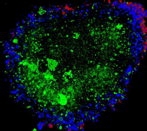
Representative imaging approach for cellular localization, morphology, and three-dimensional structural analysis.
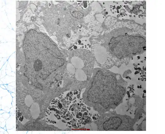
Electron microscopy can provide high-resolution information about cellular and subcellular morphology where appropriate.
The platform can support a broad range of experimental questions involving human neural development and disease biology.
Investigate cellular and structural processes involved in early human neural development and developmental patterning.
Explore how selected genetic perturbations may influence developmental phenotypes in a human-derived 3D neural system.
Study cellular mechanisms underlying neural differentiation, organization, signalling, and disease-associated changes.
Evaluate experimental compounds and investigate biological responses within a human-relevant neural model context.
Support research into cellular and structural effects associated with candidate compounds or environmental exposures.
Generate human-derived experimental data that can complement conventional cellular and preclinical research models.
Final specifications should be confirmed against the current KYAH product documentation and the requirements of the intended study.
Fetal brain organoids can be incorporated into workflows involving imaging, molecular profiling, perturbation experiments, and comparative phenotyping.
Depending on the study, researchers may combine organoid models with complementary techniques such as immunostaining, microscopy, gene-expression analysis, transcriptomics, or other validated assays.
Different studies require different endpoints. The organoid workflow can be aligned with the biological question, analytical method, and downstream research objectives.
Define model requirements, experimental groups, controls, endpoints, and analytical objectives.
Select suitable structural, cellular, molecular, or functional characterization approaches.
Discuss integration of organoid models into developmental, disease, screening, or translational workflows.
Common questions about model purpose, research applications, characterization, and experimental use.
Discuss your developmental neuroscience, disease modelling, imaging, screening, or translational research requirements with the KYAH Healthcare team.
Contact KYAH HealthcareResearch disclaimer: Fetal brain organoids are in-vitro experimental model systems and do not represent complete human fetal brain physiology. Specific biological characteristics, developmental states, assay compatibility, specifications, and performance parameters depend on the model, culture protocol, characterization method, and study design. Product-specific specifications should be confirmed with KYAH Healthcare before use in a research program.