Human Cerebral Organoids.
Three-dimensional human cerebral organoid models designed to support research into cortical development, neural biology, disease mechanisms, imaging, drug discovery, and experimental neuroscience.
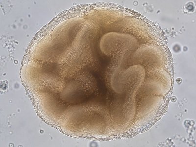
A Three-Dimensional Model of Human Cerebral Biology
Cerebral organoids are three-dimensional biological models that can reproduce selected features of human neural development and organization in an in-vitro research environment.
Compared with conventional two-dimensional neural cultures, three-dimensional models can provide additional spatial context for studying cellular organization, tissue development, morphology, and experimental responses.
Cerebral organoid systems can therefore be incorporated into research workflows investigating neurodevelopment, disease-associated mechanisms, pharmacological responses, neurotoxicity, and other areas of human neuroscience.
Cerebral Biology in a 3D Research Environment
Cerebral organoid models can provide complementary biological information for studies of human neural development, cellular organization, and experimental responses.
Cortical Development
Support investigations into developmental processes associated with neural progenitor populations, neuronal differentiation, and cortical tissue organization.
Neural Organization
Three-dimensional tissue architecture provides spatial context for examining cellular organization and morphology within developing neural models.
Cellular Characterization
Molecular and imaging-based methods can be incorporated to investigate cellular markers, morphology, and biological characteristics.
Functional Research
Cerebral organoid models can be incorporated into experimental workflows designed to investigate neural activity and functional responses.
Disease Modelling
Patient-derived or genetically modified research models may be used to investigate mechanisms associated with neurological disorders.
Drug Response Studies
Three-dimensional neural models can support experimental assessment of compound responses, toxicity, and other pharmacological endpoints.
From Model Development to Experimental Analysis
A cerebral organoid research workflow can combine model generation, culture, experimental treatment, imaging, and biological characterization.
Model Generation
Establishment of a three-dimensional cerebral organoid research model using an appropriate stem-cell-based differentiation strategy.
Culture & Maturation
Controlled culture conditions are used to support development and experimental maintenance of the three-dimensional model.
Experimental Study
Models can be incorporated into research studies involving compounds, disease-associated conditions, or defined biological questions.
Imaging & Characterization
Appropriate analytical approaches can be used to evaluate morphology, cellular features, and experimental endpoints.
Visualizing Three-Dimensional Neural Models
Imaging approaches can provide complementary information about morphology, organization, and structural features of three-dimensional neural research models.
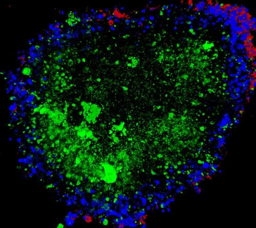
Confocal Microscopy
Confocal microscopy can support optical visualization of three-dimensional structures and cellular organization, depending on the staining and imaging workflow used.
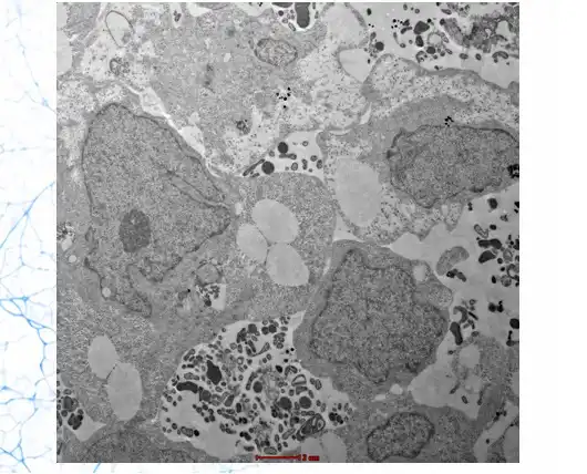
Transmission Electron Microscopy
Transmission electron microscopy can provide high-resolution ultrastructural information and may complement optical imaging approaches in biological model characterization.
Applications Across Human Neuroscience Research
Cerebral organoid models can be incorporated into a range of experimental neuroscience and pharmaceutical research workflows.
Neurodevelopment Research
Investigate selected aspects of human neural development, cellular differentiation, and cerebral tissue organization.
Neurodevelopmental Disease
Study disease-associated mechanisms using appropriate patient-derived or genetically modified research models.
Drug Discovery
Evaluate experimental compound responses using three-dimensional human neural research models.
Neurotoxicity Research
Investigate potential effects of compounds and environmental factors on developing neural model systems.
Electrophysiology Studies
Where supported by the experimental setup, cerebral organoid models can be incorporated into studies of neural electrical activity.
Imaging-Based Screening
Imaging workflows can be used to evaluate morphological and cellular changes following experimental interventions.
Research Model Considerations
The exact characteristics of a cerebral organoid model depend on the differentiation strategy, stem-cell source, maturation conditions, analytical methods, and intended experimental application.
| Model Type | Three-dimensional cerebral organoid research model |
| Biological Focus | Human cerebral and neural biology |
| Research Areas | Neurodevelopment, disease modelling, drug research, imaging and experimental neuroscience |
| Characterization | Imaging and molecular or cellular analytical approaches as appropriate to the study |
| Imaging | Confocal microscopy and other suitable imaging approaches |
| Experimental Format | To be determined according to research requirements and model configuration |
| Customization | Project-specific requirements can be discussed with the KYAH Healthcare team |
Cerebral Organoid Research FAQs
General information for researchers evaluating three-dimensional cerebral organoid models.
Cerebral organoids are three-dimensional experimental neural models that can reproduce selected aspects of human cerebral development and organization in an in-vitro setting.
Depending on the model and experimental design, they can support research into neurodevelopment, disease mechanisms, drug responses, neurotoxicity, imaging, and other areas of human neuroscience.
Yes. Cerebral organoid models can be incorporated into experimental drug discovery and response studies where the model characteristics and assay design are appropriate for the research question.
Patient-derived or genetically modified stem-cell lines may be incorporated into disease-modelling workflows where the required model development and characterization are available.
Depending on the study, researchers may use confocal microscopy, electron microscopy, fluorescence-based imaging, and other suitable analytical approaches.
Yes. Researchers can contact KYAH Healthcare to discuss model requirements, experimental objectives, characterization needs, and potential project configurations.
Build Your Cerebral Organoid Research Program
Discuss your neuroscience research objectives with KYAH Healthcare and explore suitable cerebral organoid models, characterization approaches, and project requirements.