01
Three-Dimensional Organization
Organoid models provide a three-dimensional environment for studying biological organization that can complement conventional two-dimensional cell culture approaches.
Human-relevant three-dimensional models designed to support cerebellar neuroscience research, biological characterization, and experimental disease modelling.
Cerebellar organoids are three-dimensional experimental models that can be used to investigate aspects of cerebellar biology within a structured in-vitro research system.
These models provide a research framework for studying cellular organization, developmental processes, biological mechanisms, and experimental responses relevant to cerebellar neuroscience.
KYAH Healthcare's organoid research platform brings together three-dimensional model systems, imaging, and characterization approaches to support human-relevant neuroscience research.
A three-dimensional model system can provide complementary perspectives for investigating biological organization and experimental responses.
Organoid models provide a three-dimensional environment for studying biological organization that can complement conventional two-dimensional cell culture approaches.
Cerebellar organoid systems can support experimental investigations focused on cerebellar development, organization, and biological mechanisms.
Imaging and complementary analytical approaches can be incorporated to characterize structural and cellular features of organoid models.
Cerebellar organoids can be incorporated into research workflows addressing different biological and experimental questions.
Research into cellular organization and developmental processes associated with cerebellar biology.
Investigation of cellular arrangement, morphology, and structural characteristics within three-dimensional models.
Experimental modelling of biological mechanisms associated with neurological and cerebellar disease research.
Three-dimensional research models can provide a controlled system for investigating experimental responses to candidate interventions.
Microscopy can be used to observe morphology, organization, and structural features of three-dimensional biological models.
Cerebellar organoid models can be incorporated into defined research workflows according to specific experimental objectives.
Complementary microscopy approaches can provide different levels of information for the characterization of three-dimensional biological research models.
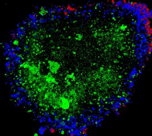
Confocal imaging can support optical visualization of three-dimensional structures and cellular organization within biological research models.
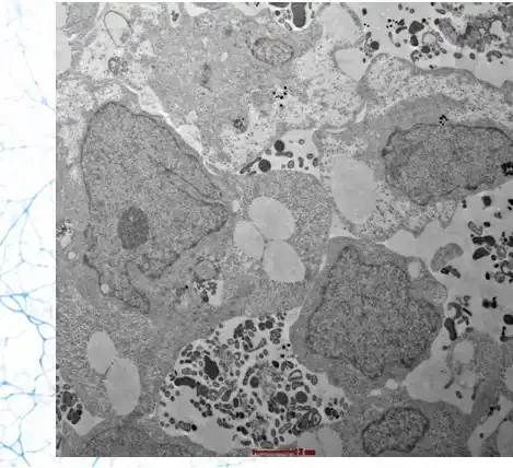
Electron microscopy can provide high-resolution structural information that complements other imaging and characterization approaches.
A cerebellar organoid research workflow can bring together model development, controlled experimental observation, imaging, and characterization.
Our approach combines three-dimensional model systems with imaging and characterization to support human-relevant neuroscience research.
Organoid-based research models.
Research-oriented human biological models.
Microscopy-supported characterization.
Designed around defined experimental objectives.
Common questions about the research role of cerebellar organoid models.
Connect with KYAH Healthcare to discuss cerebellar organoid models, characterization approaches, research requirements, and potential applications.